Pulmonary function testing measures the function of lung capacity and lung and chest wall mechanics to determine whether or not the patient has a lung problem. Pulmonary function testing is commonly referred to as “PFT” and such tests are usually performed by Certified or Registered Pulmonary Function Technologists (CPFT or RPFT) who are credentialed by the National Board for Respiratory Care (NBRC). When a patient is referred for pulmonary function testing, it means that a battery of tests may be carried-out including simple screening spirometry, static lung volume measurement, diffusing capacity for carbon monoxide, airways resistance, respiratory muscle strength, and arterial blood gases. Spirometry is the standard method for measuring most relative lung volumes; however, it is not capable of providing information about absolute volumes of air in the lung. Thus a different approach is required to measure residual volume, functional residual capacity, and total lung capacity. Two of the most common methods of obtaining information about these volumes are gas dilution tests and body plethysmography.
 Body plethysmography is a very sensitive lung measurement used to detect lung pathology that might be missed with conventional pulmonary function tests. This method of obtaining the absolute volume of air within one’s lungs may also be used in situations where several repeated trials are required or where the patient is unable to perform the multibreath tests. The technique requires moderately complex coaching and instruction for the patient.
 Ultrasound is a non-invasive procedure that used sound waves and a computer to create images of soft tissue structures such as muscles, blood vessels, and organs. It is used to diagnose a number of conditions that may not be adequately assessed with other imaging methods such as X-ray, CT, or MRI. GE Healthcare’s new compact Vivid™ ultrasound unit combines advanced imaging power in a rugged, portable, easily-cleanable, and light-weight (less than 10 pounds) unit that can be carried anywhere in the hospital or to an office-based physician practice. It is also capable of 2D transesophageal echocardiography (TEE) and intracardiac echo (ICE).
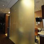 Facilitating observation without compromising patient safety and privacy is a major challenge for hospitals. Curtains are used in many areas — including viewing walls of operating rooms, sliding doors in ICUs, and for windows between patient rooms and corridors — but they can become contaminated with drug-resistant bacteria and are difficult to sterilize. A new technology playing a pivotal role in providing observation without contamination and enhancing patient privacy is liquid crystal privacy glass. This electrically activated, switchable glazing technology instantly changes from transparent to frosted white, creating 100 percent privacy. It can be activated with a power button, remote control, or a smartphone as well as automatically using light and motion sensors. When in the powered state, the panels are clear, allowing full view and daylight to pass through. When unpowered, the view is completely obscured. Privacy glass is well suited to conference rooms, partitions, hospitals, front entrance ways, sidelights, toilet and bathrooms, and windows. It can also be installed alongside existing security glass or fire resistant glass panels to ensure maximum security and patient safety.